January 2019
Transmissible spongiform encephalopathies (TSEs) are a family of chronic neurodegenerative diseases which are a human health concern and pose a threat to agricultural and natural resources. They are characterised by degeneration of brain and spinal tissue which include such diseases as Creutzfeldt Jakob Disease (CJD) in humans, bovine spongiform encephalopathy (BSE) in cattle and scrapie in sheep and goats. Classical BSE had probably been present in the UK cattle population since the 1970’s or earlier, but was initially diagnosed in cattle in 1986, subsequently developing into a European and then global animal health and food safety concern.
BSE is caused by the accumulation in of an abnormal prion protein in specified risk materials such as the eyes, brain and spinal cord. While the origin and development of BSE is still being researched, it is believed that classical BSE occurred after dietary intake by cattle of mammalian protein feed made with BSE infected animal tissues (such as meat-and-bone meal) and reached epidemic proportions in the late 1980’s and early 1990, with over 185,00 confirmed cases in cattle worldwide (more than 95% in the UK). In 2008 a case of BSE was detected in an Aberdeen, which is believed to be due to a rare spontaneous variant.
Public concern about the effect on human health arose because of the indirect evidence in 1996 linking variant Creutzfeldt-Jakob disease (vCJD) in humans with the consumption of meat from BSE-infected cattle and the inability to predict the numbers of likely vCJD cases. Because of the public concern, BSE strongly influenced medical, agricultural, economic and political issues in Europe.
Currently, as a result of the successful control measures implemented, the occurrence of classical BSE is extremely low, as is the public health risk. In the United Kingdom, Northern Ireland is considered to have a negligible BSE risk status by the World organization for Animal Health and England and Wales are considered to have controlled BSE risk status. Scotland’s negligible risk status was suspended in October 2018 following the detection of a case of BSE.
Unprecedented infectious pathogens, Transmissible Spongiform Encephalopathies (TSEs), are a class of progressive, fatal neurogenerative diseases characterised by tiny holes in the brain giving a ‘spongy’ appearance. TSEs do not usually have high morbidity or mortality in a number of mammals including humans.
Despite the low disease and mortality rate, TSEs remain important due to public health concerns, including variant Creutzfeldt-Jakob disease (CJD) identified in 1996 in Great Britain and subsequently other European countries, which may have resulted from humans eating beef from cattle that had the TSE disease bovine spongiform encephalopathy (BSE). Trade issues surrounding the movement of animals with TSEs are also important. Global commerce today is immense (over $17 trillion in 20171), with an inherent risk factor of infectious disease emergence or spread. Populations of food animals have expanded rapidly, partly due to the economic resources and desire to buy animal protein.
Animal TSE agents that have been reported are scrapie in sheep and goats; chronic wasting disease (CWD) affecting deer, elk, and moose; transmissible mink encephalopathy (TME); bovine spongiform encephalopathy (BSE) in cattle and feline spongiform encephalopathy2.
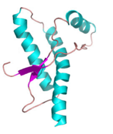
Prions are proteinaceous infectious particles which lack nucleic acid. The prion protein is an endogenous protein, occurring in most cells of all mammals but particularly produced by neurons. Cellular Prion Protein (PrPC) is a
 |
|
Image courtesy of Open-i |
protein which occurs naturally and is found in cells of the central nervous system (such as the brain and the spinal cord) and other tissues. Prion Proteins (PrP) are usually a protease-sensitive sialoglycoprotein which is anchored to membranes via glycosyl-phosphatidylinositol3.
When a PrPC molecule is altered (misfolded) through a process with another protein, it becomes infectious in the absence of nucleic acid. A portion of the prion’s α-helical and coil structure is refolded into flattened β-sheet through autolytic conversion, causing extreme changes to the physicochemical properties of the PrP. The disease-causing form of the naturally occurring prion protein, termed PrP Scrapie (PrPSc), is different to PrPC only in its tertiary and quaternary structure and has a partial resistance to proteinase K (PK, a subtilisin-related serine protease) digestion. The PK-resistant core of PrPSc is represented as Protease Resistant Protein (PrPres).
Prions are completely lacking in nucleic acid and encode strain specific properties into the tertiary structure of the PrPSc 4 by a mechanism which is not fully understood, but thought to utilise an uncharacterised intermediate conformer; the formation of infectious prions in vitro may require cellular cofactors such as polyanionic molecules. The in-vivo infectivity of prions depends on self-perpetuating properties of PrP conformers; for example, PrP amyloid fibres possess strong self-propagation capability, but do not have a strong association with infectivity. The misfolded prions then accumulate to form plaques. It is believed that prions may be self-propagating or amyloid forms of PrP; in this self-propagating form the end of the propagating fibrils are the infectious entity and fibre fragmentation causes the exponential rise in prion titre5.
PrPres varies in molecular size of the residual protein core dependent on the position of proteinase K cleavage sites. The micro heterogeneity based on differential occupancy of two N-linked glycosylation sites in PrP may also affect the molecular size. Conformational stability may vary in different prion disease types6.
Classical bovine spongiform encephalopathy (BSE) is a prion disease of domesticated cattle presenting as a slowly progressive neurological disorder. It is believed that classical BSE was first observed on a farm in West Sussex in 1984 and identified by a Central Veterinary Laboratory (CVL) pathologist as "spongiform encephalopathy" in a report dated 19 September 1985. It should be noted that, with a possible period of 2-8 years before clinical signs develop, BSE may have been entering the food and feed chain from at least as early as 1970.
The epidemic in cattle peaked in 1992 with nearly 1000 cases a week being diagnosed. Research has shown that the epidemiology and probably aetiology is heterogeneous rather than the single, epidemiologically and biologically homogeneous type. Three types (classical (C) and two atypical (H, L) types) have been identified, mainly on the basis of characteristics of the proteinase K (PK)-resistant core of the misfolded prion protein associated with the disease (PrPres). The two newer prion strains were identified in France and Italy in 2004. The types refer to the molecular weight in relations to C-type BSE; the Italian type has a lower molecular weight of unglycosylated glycoform than seen in previous BSE cases in the UK and a widespread occurrence of PrPSc-containing amyloid plaques in brain tissue, giving rise to the term L-type BSE. There is no link in atypical BSE to a common infection source and these 2 types are believed to be sporadic and may therefore never be eradicated. There is also evidence from experimental animals that atypical BSE may be able to convert to classic BSE when passaged through animals7.
The French variant with its higher molecular weight of PrPres bands compared to C-type BSE led to it being given the high or H-type BSE designation. Atypical BSE (either L or H) has since been detected (in cattle 8 years of age and older) in numerous countries, including Japan, Poland, Sweden, the United Kingdom and the United States. There are also a further two types of atypical BSE reported in Japan and Belgium that have yet to be classified. The original BSE epidemic in the UK is believed to have been caused by the inclusion in animal feed of meat and bone meal produced from animal carcasses fed to cattle, but the recent discovery of atypical types may indicate sporadic occurrence.
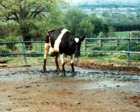
It may take between 2 and 8 years for cattle to display signs of having acquired classical BSE due to the slow development of the disease. Symptoms typically present as a change in activity level and attitude, such as nervous or aggressive
 |
|
APHIS photos by Dr. Art Davis |
behaviour. As symptoms progress, animals may show acute muscular twitching, abnormal posture, severe moaning, poor coordination, manic kicking when milked, excessive nose licking and difficulty walking or standing. Decreased milk production and weight loss may be evident despite a continuation in normal eating habits. The animal deteriorates after the presentation of clinical symptoms for up to 6 months before death. However, most animals are euthanised at the first occurrence of symptoms to confirm the BSE and prevent potential spread of the disease and the carcasses are incinerated.
The pathway of infection with BSE is unique as, other than being an entry point in the lymphatic tissues in the gut, the lymphoreticular system is not involved as the disease follows the parasympathetic and sympathetic nerve fibres of the autonomous nervous systems8.
From April 1985 to 2016 a total of 184,627 confirmed cases of BSE in cattle in Great Britain. The Bovine Spongiform Encephalopathy Order 1988 (SI 1988/1039) prohibited the sale, supply and use of certain feeding stuff for feeding to ruminants and in 1996 when new provisions came into force; these included requirements on animals exposed to BSE, the prohibition of the possession of meat and bone meal (MBM) on premises where livestock feeding stuffs were kept, the disposal and recall of MBM, and the cleansing and disinfecting of places, vehicles and equipment where MBMs had been produced, stored or used. This was more effective than The Bovine Spongiform Encephalopathy Order 1988 as it was more far reaching and did not merely address animal feed for ruminants. Since 1996 there has been a decrease in the number of BSE cases in cattle in the UK, with 0 cases confirmed on 2016. One case was confirmed on a farm in Aberdeen in 2018.
Scrapie is a fatal brain disease of sheep and goat’s endemic in many countries and recorded in the UK for well over 200 years. It is apparently naturally transmitted between sheep and has an incubation period of 2½ and 4½ years. There is no scientific evidence that scrapie can be transmitted to man either occupationally or by eating sheep meat.
While scrapie is common in sheep, recorded CJD is very rare in man. There is no evidence to suggest that the scrapie agent causes CJD through the consumption of infected sheep. Prior to 1990 it was under-diagnosed10, with the recorded annual number of UK cases fluctuating around 30. Since the creation of the UK CJD Surveillance Unit in 1990, and the intensification of surveillance, the annual totals have fluctuated around one per million of population (between 75 and 179 referrals a year to the National CJD Research & Surveillance Unit). The same surveillance procedure has yielded similar one per million annual rates in other EU countries and the rest of the world.
Variant CJD, a new form in humans, was identified in 1996, and was linked to BSE in cattle through good, although indirect, evidence.
Between 1 May 1990 and 4 November 2013 there were 3126 referrals for definite (confirmed neuropathologically) or probable vCJD cases. Of these, 176 have been confirmed or probable vCJD, all in people born before 1989. vCJD cases are believed to have reached a peak in 2000 with 27 diagnoses and 28 deaths. All vCJD clinical cases for whom genetic data are available (n=159, 90%) were met/met homozygotes at PRNP (prion-gene, pre-cursor to prion protein) codon 129 of the PrP gene11. Although the peak of the epidemic has passed, it is possible there may be future waves among different genetic groups (met/val (MV) and val/val (VV) may have longer incubation periods). In 2015, a human case of probable variant Creutzfeldt–Jakob disease was reported, based on neuropathological examination and molecular strain typing. This is the first reported case with MV genetics and may indicate a longer incubation period in people with the MV genotype.
Infectious prion proteins are far more resistant to heat than bacterial spores, with resistance to dry heat at 300°C and reports regarding the lack of prion inactivation after short-term exposure to temperatures up to 600°C 11-12. The variation in the effectiveness appears to be dependent on the nature and physical state of the infected tissues. Sterilisation with typical heat regimes does not inactivate prions, so recommendations were developed by the World Health Organisation for reprocessing equipment that may have been contaminated with TSEs in healthcare settings, where incineration is not practical (for example, equipment autoclaving), including immersion in sodium hydroxide (NaOH), heating in a gravity displacement autoclave at 121°C for 30 min, cleaning the equipment rinsing in water and then subjecting to routine sterilization13. Prion Proteins are also resistant to ionising or UV irradiation and to many chemical disinfectants including hydrogen peroxide, formaldehyde and chlorine compounds.
The CJD agent can remain infectious for 28 months at room temperature after the infected person’s death. On the other hand, pH extremes and some organic acids can inactivate prions.
BSE is typically caused when a human or animal ingests tissue which contains the prion, with young animals possibly being more susceptible to infection. Prions can accumulate in the brains of cattle after 24 months of infection. The risk from various tissue types are still not fully known, but the highest prion concentration can be found in the ileum and central nervous system.
The published oral ID50 (median infective dose where 50% of challenged cattle would become infected) for the infection of cattle with BSE is 0.2 g of BSE brain tissue, dependent on infectivity levels in the brain, which varies according to the stage of incubation (whether the infectious agent is amplifying or progressing to the central nervous system from the initially infected tissue). BSE prions have been found in the brain, spinal cord, retina and distal ileum.
Research with more sensitive techniques has also detected presence of PrPSc in adrenal glands, mesenteric lymph oesophagus, abomasum, rumen and rectum of clinically affected cattle14. vCJD15 infections have been identified in three people since December 2003. They were probably acquired from blood transfusions, although the type of prion strain may affect the ease of transmission16. The three vCJD infections followed transfusions from clinically healthy persons who then became ill more than a year after donating blood. It is therefore possible that there may be a blood-borne spread of vCJD through blood donors, who could be incubating the disease and may be a source of infection for blood transfusion recipients. The significance of a blood-borne spread of vCJD in future infections is still unknown.
Transgenic mouse bioassays are very sensitive methods in which mice are given potentially infected material and monitored for the development of disease characteristics. The test can establish whether a TSE sample is infective, the level of infectivity in the sample and the TSE strain present. The incubation period for TSEs in mice can be up to 18 months.
Immunoblot/Western Blot is a biochemical method where brain tissue is homogenised and then treated with a protease enzyme that destroys normal prion protein but not the abnormal protein. The brain sample is separated by gel electrophoresis and abnormal prion protein molecules can be detected using antibodies linked to an enzyme that results in a chemical reaction. Immunoblot tests typically take 2 to 3 days to complete and are only performed on frozen tissue. There is an optimised Western Blot approved for use by the European Union in 1999 which takes 6 to 8 hours to complete.
Immunohistochemistry (IHC) is a laboratory method of microscopic examination of brain tissue. Antibodies are linked to enzymes that show a chemical reaction that detects the abnormal form of prion protein found in BSE. This type of testing requires 2 to 3 days to complete and can be performed on fixed or frozen tissue.
ELISA (Enzyme-Linked Immunosorbent Assay) is a biochemical assay that uses antibody-antigen interactions. First, tissue is homogenised. Any normal prion protein is destroyed by a protease enzyme and the remaining abnormal prion protein is bound to the surface of a clear, microtitre well. Abnormal proteins are detected immunologically using antibodies linked to an enzyme and exposed to a chemical substrate, providing a signal in the form of a colour change or light emission, which are picked up by a spectrophotometer. Test results for BSE using the ELISA method can be confirmed through immunohistochemistry and Western Blot testing. ELISA tests for BSE can usually be completed in about 4 hours.
In histopathology, a microscope is used to detect spongiform patterns characteristic of BSE during an examination of sections of brain tissue. Histopathology tests take from 2 to 5 days to complete and are best performed on fixed tissue.
Agriculture
- Mammalian protein must not be in feed to ruminant animals. The incorporation of mammalian meat and bone meal in any farmed livestock feed is prohibited
- Except in tightly defined circumstances, mammalian meat and bone meal material must not be on premises where livestock feed is used, produced or stored
- If feed is mixed for both ruminant and non-ruminant farmed animals, and material prohibited for ruminants is used for the non-ruminant animal feed (e.g. fishmeal from non-ruminants), commingling and cross contamination of prohibited and non-prohibited materials must be avoided by following separation or clean-out procedures
- Written procedures must be maintained of feed and to provide traceability from receipt to distribution
- Separate facilities and equipment should be used for blending or storage of prohibited and non-prohibited materials e.g. separate buildings, rooms and protective clothing
- If complete feed (feed that you do not mix before feeding), is purchased, copies of all purchase invoices and labelling for feed received that contains animal protein products should be maintained. Invoices and labelling should be kept available for inspection and copying if needed
- If you suspect any animal is infected with BSE it must be reported by calling the Defra Rural Services Helpline. Prompt reporting allows time for a more thorough clinical examination, reduces the stress on the animal, reduces the risk of injury to handlers, may help to detect new strains of BSE, and safeguards public and animal health by removing potentially infected animals at an early stage.
- Effective cleaning procedures should be used on all equipment and conveyances that handle both prohibited and non-prohibited material
- When handling BSE carcasses:
-Cover cuts and abrasions with waterproof dressings before work starts
-Wear protective clothing including gloves
-Use eye protection if there is risk of splashing
-Wash your hands before eating drinking and smoking
-Wash down contaminated areas with detergent and water
-Rinse protective clothing free of debris after use, and wash with water and detergent
Slaughter and Manufacturing
- There must be separate equipment or facilities for the mixing or storage of prohibited and non-prohibited product
- Slaughter lines from ruminants should be physically separated from those for non-ruminants
- Non-ruminant blood should be collected, stored and transported in collection and storage systems which are physically separated from ruminant blood collection systems
- Train staff to ensure that all staff involved SRM management in the removal, separation, staining and disposal requirements. This should include how to avoid contaminating meat with SRM during the slaughter process
- All specified risk material (SRM) (the tissues of cattle, sheep and goats which are most likely to contain the BSE agent) must be removed in either the slaughterhouse or cutting plant
- Stain and dispose of Specified Risk Materials (SRM) in accordance with Regulations
Sources: 17,18,19, 20, 21
- World Trade Statistical Review 2017. https://www.wto.org/english/res_e/statis_e/wts2017_e/wts2017_e.pdf
- Bruce Chesebro. Introduction to the transmissible spongiform encephalopathies or prion diseases. 2003; 66(1): 1-20 doi:10.1093/bmb/66.1.1
- Stanley B. Prusiner. Prions. 1998; 95(23): 13363-13383; doi:10.1073/pnas.95.23.13363
- Dudas S, Yang J, Graham C, Czub M, McAllister TA, et al. Molecular, Biochemical and Genetic Characteristics of BSE in Canada. PLoS ONE 2010; 5(5): e10638. doi:10.1371/journal.pone.0010638
- Bernd Jansen, Christa Hilmes, Detlev Jung. Bovine Spongiform Encephalopathy and Foot-and-Mouth Disease, Two Animal Epidemics Transferable To Humans. Implications for Occupational Safety and Measures Adopted in Germany. Central European Journal of Occupational and Environmental Medicine. 2001; 7(1)
- Giles K, Glidden DV, Beckwith R, Seoanes R, Peretz D, et al. Resistance of Bovine Spongiform Encephalopathy (BSE) Prions to Inactivation. PLoS Pathog 2008; 4(11): e1000206. doi:10.1371/journal.ppat.1000206
- Sandor Dudas, Stefanie Czub, Atypical BSE: Current Knowledge and Knowledge Gaps, Food Safety, 2017, Volume 5, Issue 1, Pages 10-13, https://doi.org/10.14252/foodsafetyfscj.2016028
- Anne Balkema-Buschmann, Martin Eiden, Christine Hoffmann, Martin Kaatz, Ute Ziegler, Markus Keller and Martin H. Groschup. BSE infectivity in the absence of detectable PrPSc accumulation in the tongue and nasal mucosa of terminally diseased cattle. J Gen Virol 2011; 92:467-476
- World Organisation for Animal Health. Number of cases of bovine spongiform encephalopathy (BSE) reported in the United Kingdom. http://www.oie.int/animal-health-in-the-world/bse-specific-data/number-of-cases-in-the-united-kingdom/
- Bruton CJ, Bruton RK, Gentleman SM, Roberts GW Diagnosis and incidence of prion (Creutzfeldt-Jakob) disease: a retrospective archival survey with implications for future research. Neurodegeneration 1995; 4(4):357-68
- The National CJD Research & Surveillance Unit (NCJDRSU). http://www.cjd.ed.ac.uk/index.html
- Martin Franz et al. Detection of PrPSc in peripheral tissues of clinically affected cattle after oral challenge with bovine spongiform encephalopathy J Gen Virol 2012; 93:2740-2748. Published ahead of print 22 August 2012, doi:10.1099/vir.0.044578-0
- WTO (World Trade Organization). World trade 2008, prospects for 2009: WTO sees 9% global trade decline in 2009 as recession strike. 2009. http://www.wto.org/english/news_e/pres09_e/pr554_e.htm
- WHO Infection Control Guidelines for Transmissible Spongiform Encephalopathies. Report of a WHO Consultation Geneva, Switzerland, 23 –26 March 1999
- National Institute of Neurological Disorders and Stroke. Creutzfeldt-Jakob Disease Fact Sheet. https://www.ninds.nih.gov/Disorders/Patient-Caregiver-Education/Fact-Sheets/Creutzfeldt-Jakob-Disease-Fact-Sheet
- A General Model of Prion Strains and Their Pathogenicity John Collinge and Anthony R. Clarke. 2007; (5852), 930-936. [DOI:10.1126/science.1138718]
- Regulation (EC) No 999/2001 of the European Parliament and of the Council. Laying down rules for the prevention, control and eradication of certain transmissible spongiform encephalopathies. 22 May 2001.
http://eurlex.europa.eu/LexUriServ/LexUriServ.do?uri=OJ:L:2001:147:0001:0040:EN:PDF
- Food Standards Agency. BSE controls explained. https://www.food.gov.uk/safety-hygiene/bovine-spongiform-encephalopathy-bse - https://acss.food.gov.uk/sites/default/files/multimedia/pdfs/publication/bsenewleaflet.pdf
- Health and Safety Executive. BSE and carcass disposal. https://www.hse.gov.uk/biosafety/diseases/bovine.htm
- Meat industry guide, Food Standards Agency. https://www.food.gov.uk/business-guidance/meat-industry-guide
- Animal and Plant Health Agency. Guidance note on Feed Controls in the Transmissible Spongiform Encephalopathies Regulations. January 2018. https://assets.publishing.service.gov.uk/government/uploads/system/uploa...
Institute of Food Science & Technology has authorised the publication of the following updated Information Statement on Bovine Spongiform Encephalopathy dated January 2019, replacing that of January 2014.
This updated Information Statement has been prepared by Julie Ashmore FIFST, peer reviewed by professional members of IFST and approved by the IFST Scientific Comittee.
The Institute takes every possible care in compiling, preparing and issuing the information contained in IFST Information Statements, but can accept no liability whatsoever in connection with them. Nothing in them should be construed as absolving anyone from complying with legal requirements. They are provided for general information and guidance and to express expert professional interpretation and opinion, on important food-related issues.